Surface Analysis
XPS (X-ray Photoelectron Spectroscopy, X선 광전자 분석기)신소재융합기기센터
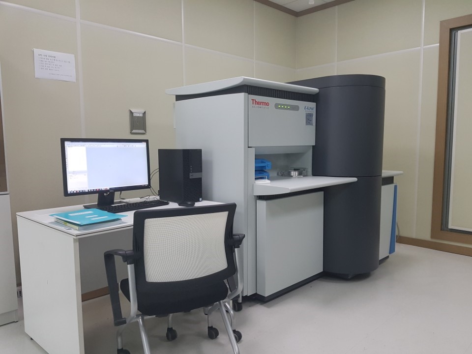
| 장비 한글명 | X선 광전자 분석기 | Equipment | XPS (X-ray Photoelectron Spectroscopy) |
|---|---|---|---|
| Model | K-alpha plus | Maker | Thermo Fisher Scientific |
| Purchase Year | 2017 | Location | Room 727, NIX |
| Operator | Ji Sook Kim | Tel | e-mail: js@suwon.ac.kr Tel.: 031-229-8665 |
| Fees | Inside University Use
Normal: 50,000 won per sample (basic five elements), 10,000 won extra charge per
additional element , Depth profile: 50,000 won per sample (basic five elements), 10,000 won extra charge per additional element Outside University Use Normal: 100,000 won per sample (basic five elements), 10,000 won extra charge per additional element, Depth profile: 150,000 won per sample (basic five elements), 10,000 won extra charge per additional element |
||
| Application Field | It is used for information about surface formation of thin-film material and bound state of
each element. It can measure composition change by thickness and analyze the amount of each composition. |
||
| Principle and Characteristic | It irradiates characteristic X-ray on solid surface in ultra high vacuum and ejects electrons in the sample. It then measures binding energy, energy level and the amount of the electron(s) in the material by measuring its kinetic energy and solidity. | ||
| Standard Features |
Range Of motion: 100 - 4000 eV X-Ray Monochromator: Al Ka Micro-focused X-Ray Spot Size: 30 - 400 µm in 5 µm steps Sampling Area: 60x60 mm Thickness (Metric) Max. Sample: 20 mm |
||
AFM (Atomic Force Microscopy, 원자힘 현미경)신소재융합기기센터
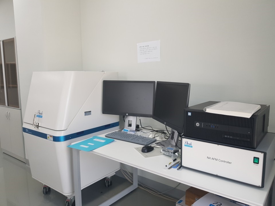
| 장비 한글명 | 원자힘 현미경 | Equipment | AFM (Atomic Force Microscopy) |
|---|---|---|---|
| Model | NX10 | Maker | Park systems |
| Purchase Year | 2017 | Location | Room 719, NIX |
| Operator | Ji Sook Kim | Tel | e-mail: js@suwon.ac.kr Tel.: 031-229-8665 |
| Fees | Inside University Use: 20,000 won per hour (Tips extra ) Outside University Use: 50,000 won per hour (Tips extra) |
||
| Application Field | 3D Analysis of Fine Surface Shape of the Sample Measurement of Magnetic and Electric Shape through MFM, EFM |
||
| Principle and Characteristics | It measures the shape and mechanical and physical characteristics of surface of the material by using gravitation and repulsive force. | ||
| Standard Features |
XY Scanner 50 µm × 50 µm (optional 10 µm × 10 µm or 100 µm × 100 µm) Resolution: 0.05 nm Position detector noise: < 0.25 nm (bandwidth: 1 kHz) Out-of-plane motion: < 2 nm (over 40 µm scan) |
||
Confocal Raman Microscopy (라만 형광 현미경)신소재융합기기센터
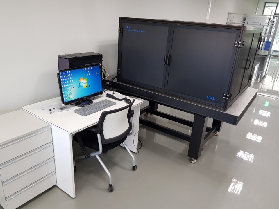
| 장비 한글명 | 라만 형광 현미경 | Equipment | Confocal Raman Microscopy |
|---|---|---|---|
| Model | FEX | Maker | Nost |
| Purchase Year | 2017 | Location | Room 718, NIX |
| Operator | Ji Sook Kim | Tel | e-mail: js@suwon.ac.kr Tel.: 031-229-8665 |
| Fees | Inside University Use: 10,000 won per point (10,000 won extra charge per additional hour) Outside University Use: 20,000 won per point (10,000 won extra charge per additional hour) |
||
| Application Field | It is used for Raman imaging and analysis, Fluorescence imaging, and dark-field imaging. It Is also used for measurement of PL. |
||
| Principle and Characteristics | When the energy larger than molecule vibration energy is given, Raman distraction is produced as a result of energy change through interaction of molecules. It enables the analysis of structure and components of molecules because the vibration energy of a molecule is distinct. | ||
| Standard Features |
Laser module Multiple excitation lasers Power control: Includes 11 steps motorized ND filter controller Laser filter selector: Motorized 3-positioning laser filter selector < 1㎛ x, y spatial resolution and < 2㎛ depth resolution with true confocal design. |
||